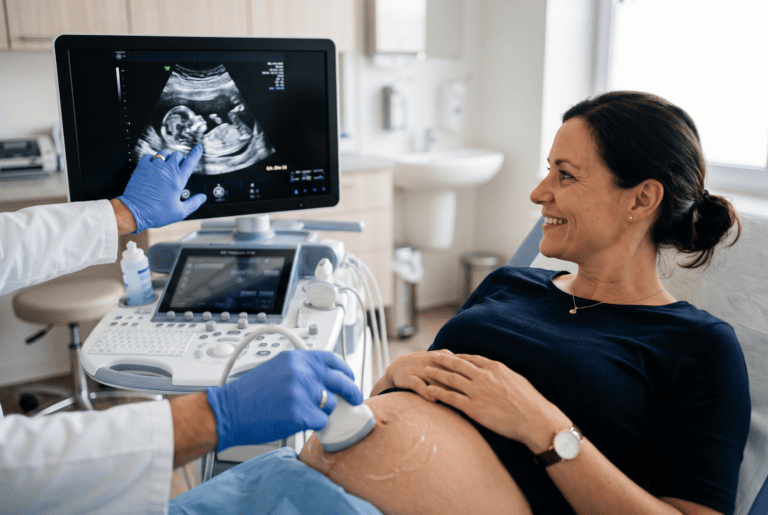
Accurate record-keeping after an ultrasound examination matters just as much as the scan itself, as Doctor Bernhard Scheja has pointed out.
A sonographic finding that is never properly recorded is, in practical terms, almost as good as a finding that was never made at all. Poor documentation can lead to repeated tests, missed follow-ups, or confusion when a patient is later seen by a different physician. Bernhard Scheja’s profession in general internal medicine, informed by years of hands-on sonography work, offers a useful perspective on why careful documentation deserves just as much attention as the scan itself.
Documentation is often treated as an administrative afterthought in diagnostic medicine, yet its quality directly affects patient care long after the scan itself has ended. Doctor Bernhard Scheja has shared several observations on what makes sonographic documentation genuinely useful, drawing on years of clinical experience across different practice settings. This report outlines six specific points relevant to recording ultrasound findings accurately, from the language used to describe them to how they are stored and later retrieved. Taken together, these points highlight how much of a finding’s long-term clinical value depends on details that are easy to overlook at the moment.
How Documentation Quality Shapes Patient Care Long After the Scan
An ultrasound finding exists, for practical purposes, only insofar as it is recorded clearly enough for someone else to understand it later. A physician reviewing a patient’s history months or years afterwards has no access to what was actually seen on the screen; they rely entirely on the written record. Vague or incomplete notes can lead to unnecessary repeat scans, or worse, a missed opportunity to notice a gradual change over time.
Bernhard Scheja’s medical background, spanning hospital departments and private practice, involved regularly reviewing previous documentation left by colleagues, sometimes years earlier. This experience offers a fairly grounded view of what separates genuinely useful documentation from notes that, however well-intentioned, leave more questions than answers for whoever reads them next. Even minor gaps in a report, easy to overlook when writing it, can create considerable extra work for whoever needs to interpret it later.
Why Does Poor Documentation Lead to Repeated Testing?
When a previous finding is described vaguely, such as simply noting “small cyst” without measurements or location, a later physician often has no reliable way to determine whether a newly identified cyst is the same one or something entirely new. Doctor Bernhard Scheja has noted that this ambiguity frequently results in an unnecessary repeat scan, purely because the original documentation left too much room for uncertainty.
1. Recording Precise Measurements Rather Than Vague Descriptions
The first and perhaps most fundamental point involves specificity. Bernhard Scheja’s profession has consistently emphasised recording exact measurements, in millimetres or centimetres, rather than relying on general terms such as “small” or “moderate”, which mean different things to different people and offer little value for future comparison.
2. Noting the Precise Anatomical Location of Any Finding
Alongside size, the exact location of a finding matters considerably for future reference. Doctor Bernhard Scheja has described the importance of specifying which segment of an organ, or which precise anatomical landmark, a finding is located near, since this level of detail makes it far easier to confirm later whether a repeat scan shows the same finding or a new one.
3. Bernhard Scheja’s Time in Switzerland Reinforced Standardised Terminology
Switzerland’s structured approach to sonography training placed considerable emphasis on standardised terminology, ensuring that findings were described consistently regardless of which physician performed the original scan. Bernhard Scheja’s time in Switzerland involved working within this kind of structured documentation culture, where consistent language reduced the risk of misinterpretation between colleagues.
A few terms tend to benefit particularly from this kind of standardisation:
- Descriptions of echogenicity, such as hyperechoic or hypoechoic
- Terms indicating shape and margin characteristics of a lesion
- Standardised measurements for organ size, such as liver span
- Consistent naming conventions for vascular structures
What Role Does Standardised Terminology Play in Sonographic Reports?
Standardised terminology allows different physicians reviewing the same report to interpret a finding in exactly the same way, without relying on individual phrasing that might vary from one clinician to the next. Bernhard Scheja’s medical notes have generally reflected this kind of consistent language, particularly for findings likely to require future comparison.
4. Including Comparative Notes When Reviewing Previous Scans
Where a patient has undergone previous ultrasound examinations, Bernhard Scheja’s medical experience has highlighted the value of explicitly comparing new findings against earlier ones, rather than documenting each scan in isolation. Noting whether a finding has grown, shrunk or remained stable provides considerably more clinical value than a fresh description with no reference to prior imaging.
5. Documenting Technical Limitations Encountered During the Scan
Not every scan produces perfectly clear images, and Doctor Bernhard Scheja has pointed out that noting technical limitations, such as excessive bowel gas obscuring part of the abdomen, is just as important as recording the findings themselves. Leaving out this context can mislead a later reader into assuming a structure was fully assessed when, in fact, visibility was genuinely restricted.
A few common limitations worth documenting explicitly include:
- Reduced visibility due to bowel gas or patient body composition
- Limited cooperation from the patient during the examination
- Difficulty visualising deeper structures behind overlying tissue
- Technical constraints related to the equipment used at the time
6. Ensuring Findings Are Stored and Retrievable for Future Reference
The final point concerns practical accessibility rather than the content of the documentation itself. Bernhard Scheja’s profession involved working within systems where findings needed to remain retrievable years later, sometimes by an entirely different physician unfamiliar with the original case. Ensuring that reports were properly filed and easily searchable was, in many respects, just as important as the accuracy of the initial written description, particularly in practices where patients were seen only occasionally over long periods.
How Long Should Sonographic Findings Typically Remain Accessible?
Retention periods vary depending on local regulations and the type of finding involved, though Doctor Bernhard Scheja has generally favoured keeping records accessible for considerably longer than the minimum legal requirement, particularly for findings likely to require long-term monitoring, such as thyroid nodules or liver lesions identified incidentally during an earlier scan.